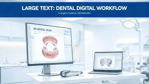
Before we get into the mechanics, here's the conclusion: an intraoral scanner works by projecting structured light onto the teeth, capturing the reflected pattern with camera sensors, and using software to stitch those frames into a 3D model. But the scanner hardware is the easy part. In the last four years, I've personally documented 14 scanning mistakes that cost our practice roughly $19,000 in remakes and delays—and every single one was a workflow problem, not a scanner hardware failure. If you understand how the software processes the scan, you'll avoid the same errors.
I manage digital implant workflows. I'm not a radiologist, and I'm not an optical engineer. I'm the person who had to explain to a lab why a crown didn't fit. That's why this article is not a spec sheet review. It's a “how does an intraoral scanner work” answer with the failures included.
The Short Answer: What the Scanner Actually Does
An intraoral scanner is a camera system with a structured light source. The wand projects a pattern—often blue laser light or LED—onto the teeth. The camera captures how that pattern deforms over the surface. Software calculates depth from the distortion, then stitches thousands of frames into a continuous 3D mesh.
That's it. The magic is in the matching.
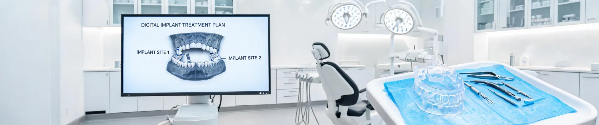
For implant cases, the scanner needs a reference. That's what a scan body is for. You screw a scan body into the implant, scan it, and the software maps its geometry to your implant library. In our practice, that's usually a dental implant Straumann case in one of the SLActive or BLT configurations. Once the scan body is recognized, the software knows the implant's exact position and angle. If the scan body is loose, or if the software misidentifies it, the whole case is compromised.
When that data goes into a CAD environment and connects with Straumann digital solutions, you can plan the case, design the surgical guide, and mill the restoration. For a single implant crown, the software matches the scan body position to the implant library, then designs a custom abutment—often on a Straumann TiBase—and the final restoration. For a guided surgery case, the same scan data supports the design of a surgical guide, so the planned implant position is carried into the operatory.
That's the vision. But the vision only works if the scan is clean.
My Initial Mistake: I Compared Scanners Like Hardware
When I first brought a scanner into our practice in 2017, I evaluated it the same way I evaluated a surgical stapler or a patient lift for our surgical suite. Tug it, turn it on, check the price. Those are mechanical tools. They do their job if the build quality is solid.
An intraoral scanner is not that kind of product. It's a software instrument with a handle attached. Two scanners with the same camera sensor can produce different results because the stitching algorithm is different. I didn't get that at first.
Here's the thing: if you choose a scanner based only on camera specs and price, you're missing the part that determines clinical success. The software workflow is the device. Everything else is a peripheral.
What the Spec Sheet Doesn't Tell You
What most people don't realize is that the accuracy number on the brochure is measured on a typodont under controlled light, with a dry arch and an operator who knows the scan path. In a real mouth, with saliva, movement, and posterior access, the working accuracy is lower. I'm not saying modern scanners are inaccurate—I'm saying that number is a ceiling, not a guarantee.
Per FTC guidelines (ftc.gov), product claims have to be truthful and substantiated. So when a rep says “this scanner is accurate to five microns,” ask for the test method. I wish I had asked sooner.
The Mistakes That Cost Us $19,000
Digital workflows were supposed to replace physical shipping. For the most part, they did. As of January 2025, USPS First-Class Mail letter postage is $0.73 (usps.com), so the price of mailing a stone model is not the real issue. The real issue is that a bad digital scan costs a lot more than postage.
I don't keep the mistakes list to feel bad about myself. I keep it because it caught 47 potential errors in the last 18 months. Here are the patterns:
1. I approved a scan that “looked fine”
In my first year, I submitted a full-arch scan with a missed margin on a premolar. The model looked clean on the screen. The lab milled the crown anyway, and it came back with a visible gap. $2,100 and a week of delay, straight to the trash. That's when I learned that a scanner can produce a beautiful model of the wrong geometry.
I want to say the first scan I ever approved took 20 minutes and looked perfect on screen. The lab still rejected it. Don't quote me on the exact time—it would've felt longer if I'd known what was coming.
2. I trusted the wrong scan body position
In September 2022, we had an implant case where the scan body was not fully seated. I didn't check it clinically before sending the file. The CAD software designed a restoration that looked perfect in the virtual model. On the day of delivery, it didn't seat. The remake cost $340 and a patient's confidence.
3. I ignored the stitching artifact
After the third rejection in Q1 2024, I created our pre-scan checklist. One item on that list: review the raw scan for “jumps” in the mesh. A stitching artifact can happen when the wand lifts off the surface too suddenly. Sometimes the software will warn you. Sometimes it will quietly create a step that you can't see unless you rotate the model.
That checklist has caught 47 potential errors since then. Not one of them was a scanner that “didn't work.” Every one was an operator or workflow issue.
How to Actually Get a Good Scan
If you're asking “how does an intraoral scanner work,” you might assume the answer is technical. It is, in part. But the practical answer is simpler: a good scan depends on a consistent path, good isolation, and knowing when to pause and rescan.
- Dry the area. Saliva creates artifacts.
- Follow the recommended scanning path. It matters more than speed.
- Check the mesh before you export. Rotate it in every dimension.
- Verify the scan body is fully seated before you scan. Always.
Not a glamorous answer, but it's the honest one.
I'd rather spend ten minutes explaining scan bodies and stitching artifacts than deal with mismatched expectations later. An informed customer asks better questions and makes faster decisions. That applies to the dentist in the chair too.
What I'd Tell a First-Time Buyer
It took me three years and maybe 300 scans to understand that the scanner is not the bottleneck. The operator is. That was humbling.
If a dental implant Straumann case with a digital workflow is your goal, buy a scanner that connects cleanly with Straumann digital solutions. Check the open architecture. Ask about the scan body library. Ask how the manufacturer handles software updates. And if you can, use a demo scanner on a patient before you buy, not just on a model.
Honestly, I'm not sure why some scanner software doesn't flag critical stitching gaps more aggressively. My best guess is that it's a design choice: the algorithm wants to feel fast and seamless. But a red warning would have saved me $2,100.
One Caveat
Modern intraoral scanners are excellent for single-unit and short-span implant restorations. For fully edentulous arches with mobile tissue, I still want a prosthodontist's input before trusting the scan. That gets into tissue compression and border molding territory, which isn't my expertise. I'd recommend consulting someone who does it daily.
The scanner is not a formula for clinical certainty. It's a tool. The real value comes from the people, the software, and the workflow around it.
If you get that wrong, no sensor in the world will save you. I know because I've paid to learn it.